Gallstones on ultrasound

these [shows me ultrasound images of his gallbladder, full of stones.

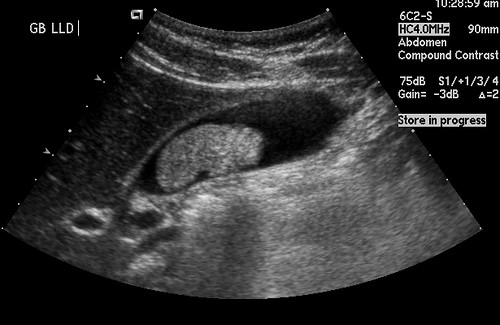
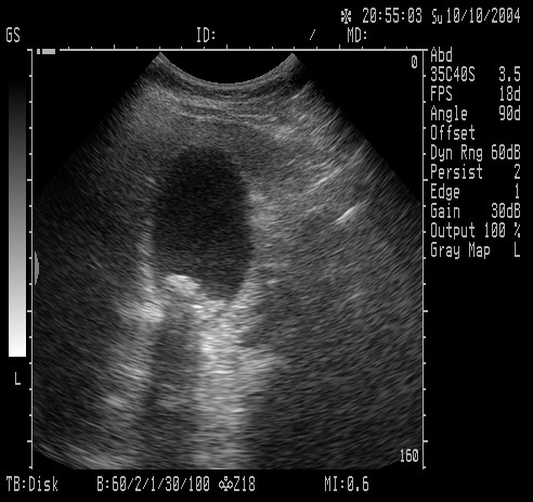
Figure 1 shows a normal gallbladder on ultrasound. Figure 2 shows a large

An ultrasound screen shows a patient's kidney. (Photograph by Brownie Harris

Ultrasound image of gallstones

I had an ultrasound yesterday. They want to see if my abdominal pains are

Ultrasonography of gallstones; A, ultrasound probe postioning; B,

The ultrasound only shows gallstones within the g.

Gallbladder or hebatobiliary ultrasound for gallstones,

1cm solitary gallstone on ultrasound. Note the acoustic shadow.

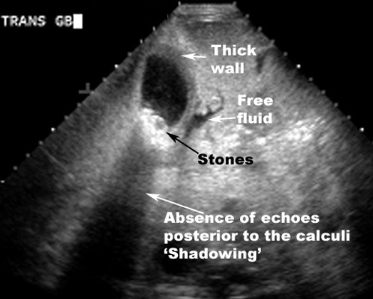
Thick GB wall; Stones in GB; Absence of echoes posterior to the calculi

Abdominal ultrasound demonstrates a gallstone lodged within the cystic duct

Figure 3: Hepatobiliary ultrasound showing multiple gallstones and

Abdominal ultrasound demonstrates a gallstone lodged within the cystic duct

Gallstones may also be discovered upon ultrasound or abdominal CT study.

Gallbladder or hebatobiliary ultrasound for gallstones,

Gallbladder carcinoma carcinoma ultrasonography and ultrasound liver

Ultrasonography gallbladder polyp stone and sludge ultrasound échographie

Gallstones on ultrasound

gallstone. Ultrasound – 1) calculi 2) gallbladder wall thickening 3) cystic
